Shilajit Natures Rejuvenator: Why I Added This Ancient Resin to My Daily Routine
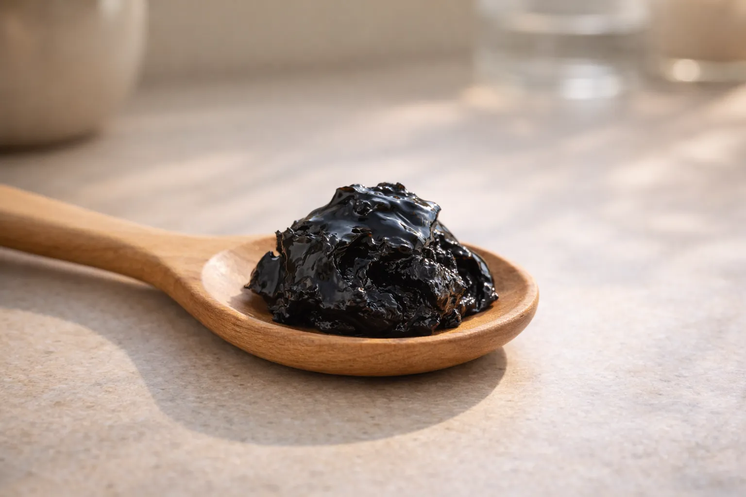
I first came across shilajit during a phase when I felt permanently drained. I slept enough, ate well, and still…
I first came across shilajit during a phase when I felt permanently drained. I slept enough, ate well, and still…
If you spend even a little time searching for trending entertainment news, TV show updates, viral social media topics, or…
Feeling overwhelmed by stress, struggling to get a good night’s sleep, or searching for a natural way to boost your…
The first time I noticed my energy dipping, I blamed stress, work, and not sleeping enough. Still, something felt off.…
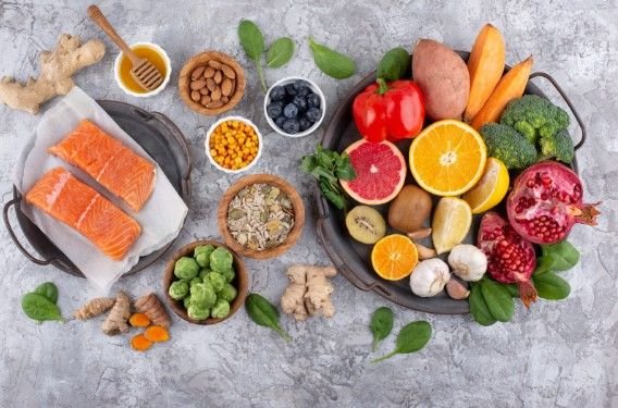
Managing acid reflux involves choosing foods that help soothe your digestive system and promote overall gut health. By making the…
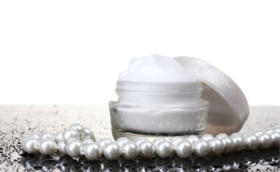
Pearl powder and extract have long been celebrated for their powerful effects on both health and beauty. With a rich…
The first time I heard someone casually ask vitamin b17 apricot kernals anyone, I was half-asleep and scrolling through wellness…
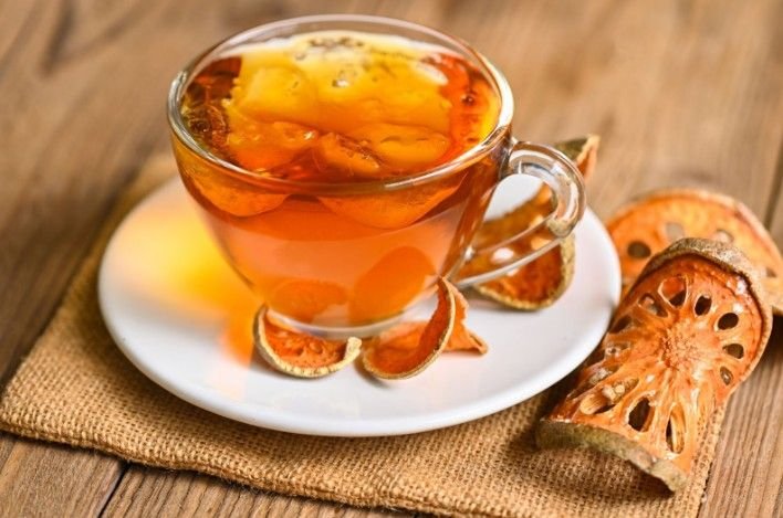
Looking for a natural, caffeine-free drink packed with health benefits? Persimmon leaf tea, known as kakinoha-cha in Japan and gamnip-cha…
I first noticed my legs feeling heavy and tired at the end of long days, especially after sitting too much…
Choosing the right paint color for your home can feel like a daunting task. I’ve been there—staring at endless paint…